Reproduction
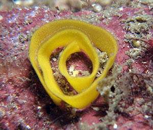
A defining characteristic of most nudibranch species is the ephemeral life style of the animals. Most members of order Nudibranchia are semelparous, meaning the adults undergo a single spawning period and die (Todd, 1981). As most nudibranchs only live for about a year, reaching maturity in a matter of days or weeks, this window for reproduction isn't very large (Rudman, 2000). Because of the short nature of their life spans, mating is opportunistic; occurring whenever mature nudibranchs come in contact with one anther (Todd, 1981). The genital opening of all nudibranchs are located on the right side of the body. During copulation the mating individuals align head to tail, with right side in proximity. Once the genital papillae of the individuals touch, the penis enters the female duct of the other (Behrens, 2005). At this point nudibranchs exchange sperm packets, which are stored in a seminal receptacle until such a time that environmental conditions are suitable for fertilization (Todd, 1981). Individuals are then capable of depositing their spawn mass on suitable substrate, spiraling in a characteristic counter-clockwise egg ribbon (Rudman, 2000). The larval state that emerges from the egg ribbons a free living veliger larva(Rudman, 2000).
|
| Figure 1. Counter-clockwise spiral egg ribbon (Picture from http://www.nudibranch.com.au) |
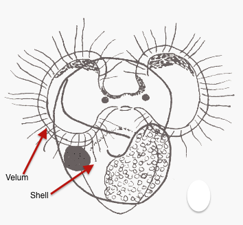
These veliger larvae are named for their swimming organ, the velum, which consists of two large, semicircular, ciliated lobes that beat the water for movement. The veliger larva has a hard, protective shell, the shape of this shell varies between species and is often used as a distinguishing characteristic in identifying genera (Todd, 1991). The longcilia of the velum function not only in locomotion, but also in suspension feeding by bringing plankton in contact with the shorted cilia of the subvelar food groove (Ruppert
et al, 2004). These larvae swim and drift in the current, sometimes covering hundreds of kilometers until they find a suitable site to anchor and undergo metamorphosis. Site selection is crucial because nudibranch species are specialized for life in a specific habitat, and are unlikely to survive in a hostile or new environment (Ruppert
et al, 2004).
|
| Figure 2. Veliger larvae (drawing by Micheal G. Hadfield) |
Feeding
All nudibranchs feed by way of a radula- a flexible, tongue-like mouthpart with rows of microscopic back pointing teeth located in the mouth (Behrens, 2005). The radula is manipulated with a series of extendable protractor muscles to act like a rasp; scraping across the substrate tearing off particles of algae or sponge. The food particles are moved by conveyer belt action back to the esophagus where it is ground into a soupy consistency for digestion (Rudman, 2000). The structure and organization of the radula’s teeth change between groups, depending on the diet of the nudibranch.
Hydroids, such as Tubularia are the preferred prey item of most Aeolids (Behrens, 2005). These are primarily small branching hydroids that provide a lot of biomass for the nudibranchs, who often pick a single feeding site and stay there for the remainder of their lifespan (Behrens, 2005).
Godiva is composed of more mobile species that actively search out these hydroid colonies with the use of their chemosensory organs, the rhinophore. This is a highly specialized sensory organ used to sense the chemical signals in the water column around them (Rudman, 2000). They use these rhinophores to hone in on the signals of their prey while their oral tentacles feel out the surrounding. The rhinophores of godiva are specialized with additional wrinkles at the distal ends to increase surface area for chemical interaction (Behrens, 2005).
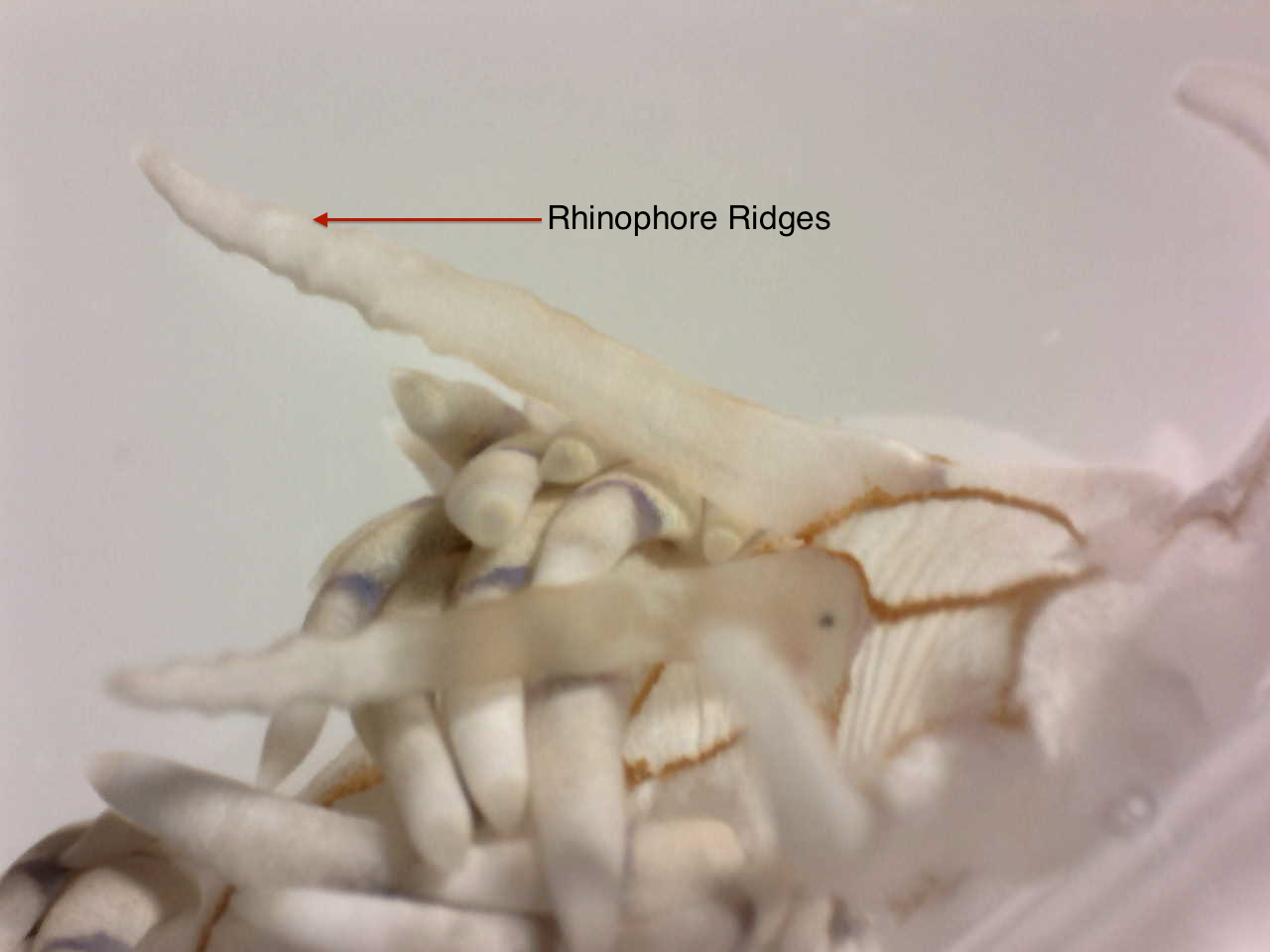 Figure 3. Rhinophores of Godiva specimen with characteristic ridges Figure 3. Rhinophores of Godiva specimen with characteristic ridges |
Excretion
Nudibranch’s expel waste through the anus, located two-thirds of the way down the right side of the body. Digestion occurs in the stomach and intestines through a series of digestive glands. Waste products are expelled from the anal pore on the right lateral side of the animal (Behrens, 2005).
Respiration
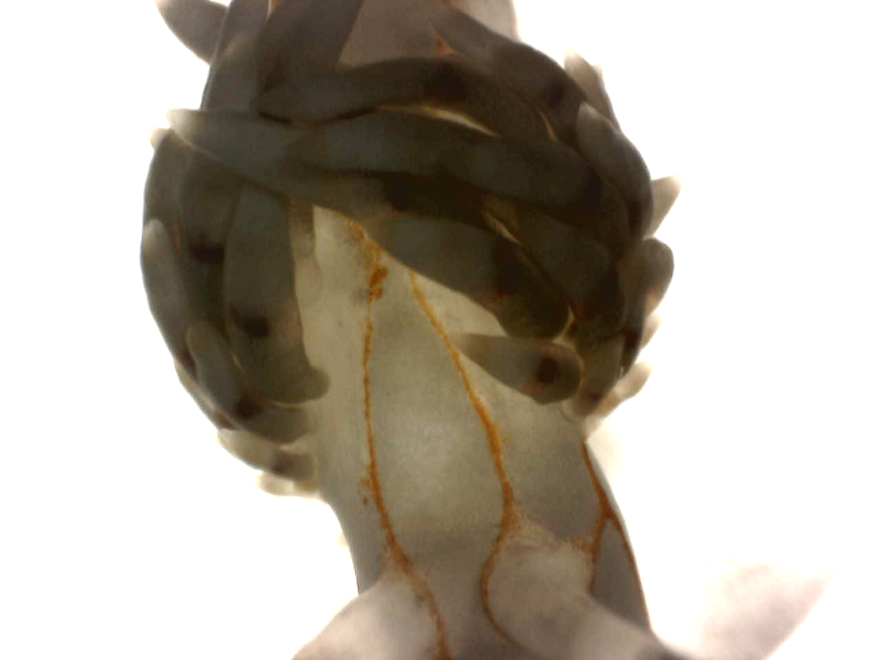
All nudibranchs exchange gas directly though the tissue ofexternal respiratory structures called “naked gills” (Behrens, 2005). Each subgroup of nudibranchs has evolved their own way of using these structures for acquiring oxygen. Aeolids use unique cigar or finger-shaped body extensions called cerata (Rudman, 2000). These cerata, singular form ceras, are thin walled, elongated sacs that provide a direct route for blood to absorb oxygen from the surrounding seawater (Rudman, 2000). Cerata are a defining characteristic of the suborder and are often quite colorful.
|
| Figure 4. Dorsal cerata complete with cnidosac tips and naked gill tissue |
Locomotion
Unlike some species of nudibranch which can use their foot to swirl and swim in the water, members of Godiva are limited to crawling along the substrate. They use their flat, flexible “foot” on the underside of the body to pull themselves along. The foot is composed of two distinct muscular parts; the thick, skirt-like outer muscle that remains in constant contact withthe substrate, an the elongated central section that makes contact with thesubstrate in muscular contractions. The latter portion is termed the “sole” andis the primary mechanism of movement in most nudibranchs. The sole also contains specialized glands for secreting mucus, which forms a thin slimy layer for the nudibranch to slide across (Behrens, 2005). The encircling outer muscleis covered fine cilia that beat to help the animal move over its slime trail.This outer portion of the foot is larger in smaller nudibranchs who are more reliant of the cilia for motion. Godiva fall into this category, being dwarfed by some of the larger sea hares.
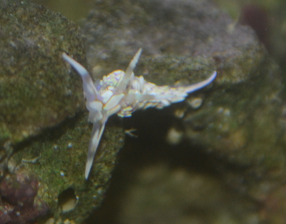
When members of this species were released into the water in a lab setting they balled themselves up, extending their cerata, and drifted to the bottom of the tank. They made no move to swim, leading to the conclusion that their singular mode of transport is crawling.
|
| Figure 5. Godiva specimen traversing boulder in lab aquarium |
Rearing
Rearing is a behavior observed in this species when specimens came in contact with irritating or foreign substances such asstinging corals or probes. The individual lifts the upper portion of its body off the substrate, extending, and waving its oral tentacles and rhinophores through the water. This behavior is likely used to adjust the position of the body to better sense food or potential threats (Behrens, 2005).